View in
3D
Speed: around
5
minutes
High resolution: less than
3
microns
What is optical coherence tomography?
OCT is an imaging techniques that provides an automatic, high-resolution scan of the ocular structures, generally of the posterior pole although its use has been extended over recent years to include the anterior segment by adapting lenses or special software, and the design of specific equipment. This provides an in vivo view with micrometric precision of the cornea, the anterior chamber, the iris, and the crystalline lens.
What does anterior OCT involve?

It is a minimally invasive, painless tests, as it involves no contact with the eye. It is based on the emitting of an infra-red light that, when reflected on the structures of the anterior segment, produces a 3D cross-cut map of the tissue.
To do so, modern equipment uses different types of technology: Time Domain (TD), Spectral Domain (SD) –with greater definition and scanning speed– and the new Swept Source, which guarantees clear photographs even in the peripheral of the cornea and in-depth images of the anterior chamber.
How is it performed?
The anterior OCT is a very fast test that is performed in the consulting room (around 5 minutes), which may or may not require pupil dilation depending on each case and on the equipment used. When the diameter of the pupil must be increased, eye drops (mydriatic drops) are used that take effect in around 15 minutes and lead to blurred vision and dazzle, which disappear after a few hours.
4 keys to anterior segment OCT
- It is a painless test, there is no contact with the eye.
- No dilation is necessary (in some cases it must be performed without dilation).
- Contact lenses do not have to be removed for any time beforehand.
- The test lasts around 10 minutes.
In what cases is it used?
Anterior optical coherence tomography detects very subtle morphological changes to the ocular structures, providing extremely valuable information beyond examinations using a slit lamp.
This test provides an objective assessment of aspects such as the thickness of the cornea in patients with keratoconus or candidates for refractive surgery; the dimensions of the anterior chamber for intraocular lens implantation, or the iridocorneal angle (forming the cornea with the iris) in cases of closed-angle glaucoma.
Associated treatments
You may be interested in
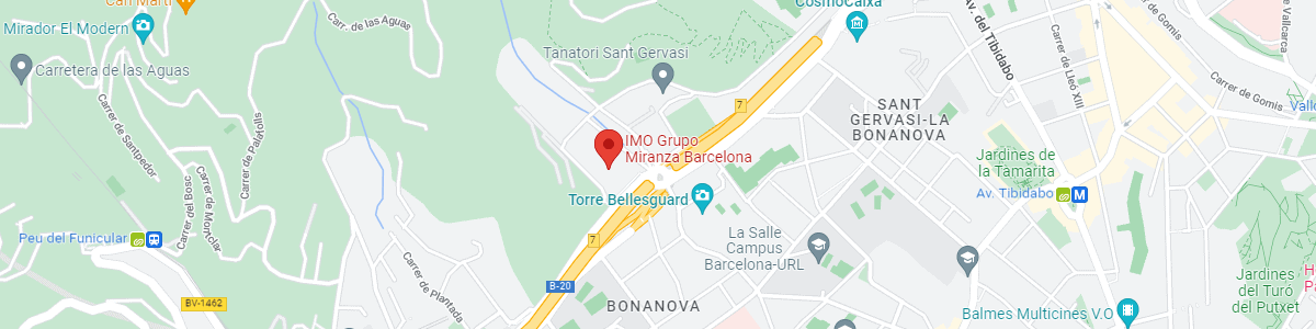
IMO Institute of Ocular Microsurgery
Josep María Lladó, 3
08035 Barcelona
Phone: (+34) 934 000 700
E-mail: international@imo.es
See map on Google Maps
By car
GPS navigator coordinates:
41º 24’ 38” N – 02º 07’ 29” E
Exit 7 of the Ronda de Dalt (mountain side). The clinic has a car park with more than 200 parking spaces.
By bus
Autobus H2: Rotonda de Bellesguard, parada 1540
Autobus 196: Josep Maria Lladó-Bellesguard, parada 3191
Autobuses H2, 123, 196: Ronda de Dalt – Bellesguard, parada 0071
How to arrive at IMO from:
IMO Madrid
C/ Valle de Pinares Llanos, 3
28035 Madrid
Phone: (+34) 910 783 783
See map in Google Maps
Public transport
Metro Lacoma (líne 7)
Autobuses:
- Lines 49 & 64, stop “Senda del Infante”
- Line N21, stop “Metro Lacoma”
Timetables
Patient care:
Monday to Friday, 8 a.m. to 8 p.m.
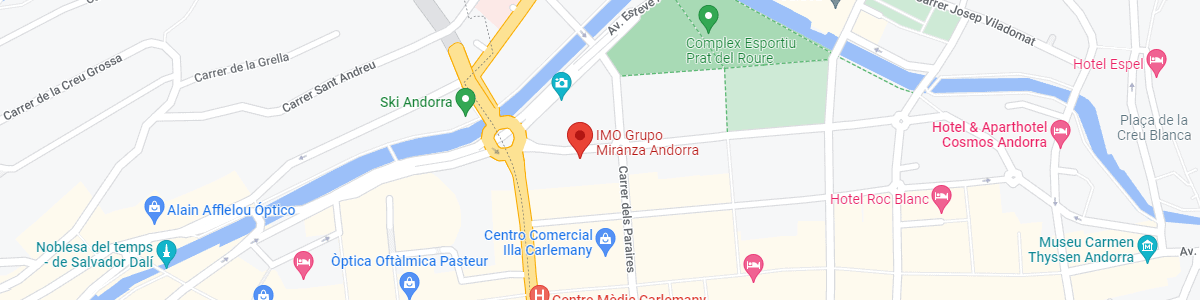
IMO Andorra
Av. de les Nacions Unides, 17
AD700 Escaldes-Engordany, Andorra
Phone: (+376) 688 55 44
See map in Google Maps
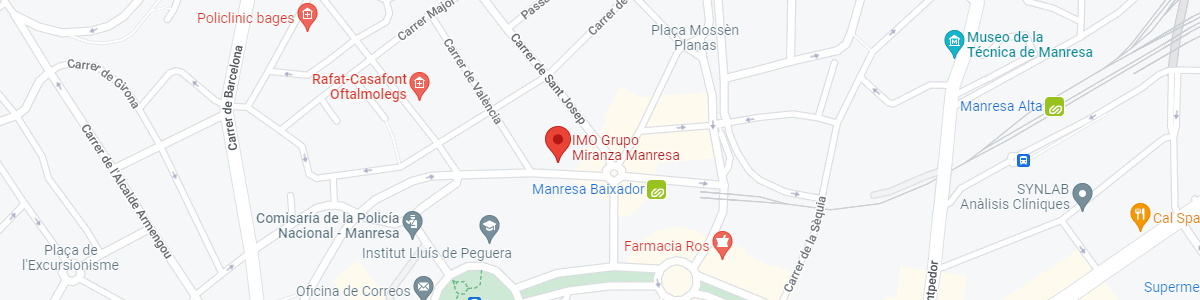
IMO Manresa
C/ Carrasco i Formiguera, 33 (Baixos)
08242 – Manresa
Tel: (+34) 938 749 160
See map in Google Maps
Public transport
FGC. Line R5 & R50 direction Manresa. Station/Stop: Baixador de Manresa
Timetables
Monday to Friday, 08:30 A.M – 13:30 PM / 15:00 PM – 20:00 PM