IMO has
3
different pieces of equipments
Images captured within
3
seconds
Duration:
3-5
minutes
What is an OCT-angio?
An OCT-angio is innovative new diagnosis equipment that uses optical coherence tomography (OCT) technology to obtain high-quality images of retinal circulation and provide a 3D view of the structures of the eye’s posterior pole.
This allows for extremely precise examination of the eye’s vascular network at different depths, showing the mobility of blood flow in the vessels in specific layers of the retinal and the choroid (the layer just below), as well as in the optic nerve head.
Of what does it consist?
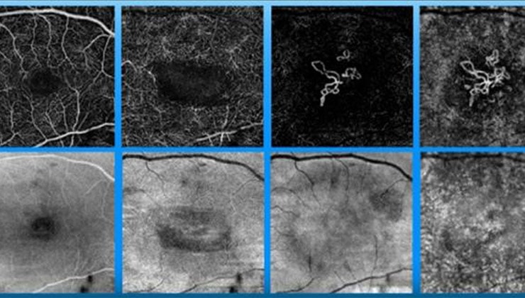
Based on OCT (light emission) technology, this technique captures images within just 3 seconds. Unlike the conventional fluorescein angiography -a test traditionally used to study retinal circulation-, it requires no intravenous injection of contrast to dye and view the vascular section.
As a result, not only is it faster to perform, but it also avoids discomfort and possible adverse effects for patients. This makes it a minimally invasive option that can be repeated more often for thorough control of diseases.
How is it performed?
The OCT-angio is performed in the consulting room in less than 10 seconds, without the need to prepare the patient beforehand or to dilate the pupil.
6 keys to the OCT-Angio
- Painless test for patients.
- Can be performed without prior preparation and without dilating the pupil.
- Can be performed on patients of any age.
- An appliance is used that emits a light into the eye through the pupil in order to analyse the different layers of the retina and the choroid.
- It is a minimally invasive test for patients.
- Test duration: 3-5 minutes.
In what cases is it used?
Included as state-of-the -art equipment in IMO consulting offices since late 2010, the OCT-angio is particularly relevant with regard to vascular diseases of the retina, as it allows for the study of abnormalities such as microaneurysms (dilation of blood capillaries) in diabetic retinopathy. Another of its main indications is AMD, as it can detect the presence of neovessels (new abnormal blood vessels).
Because this is a new technique, however, the images obtained cover just the central part of the retina and, in the case of peripheral disorders, the conventional fluorescein angiography is still required.
It is also important to remember that, as well as being a powerful diagnosis weapon, the OCT-angio is great help in monitoring and evaluating the patient’s response to treatment. The usefulness and contribution of this innovative technology to glaucoma is also being considered.
You may be interested in
IMO Institute of Ocular Microsurgery
Josep María Lladó, 3
08035 Barcelona
Phone: (+34) 934 000 700
E-mail: international@imo.es
See map on Google Maps
By car
GPS navigator coordinates:
41º 24’ 38” N – 02º 07’ 29” E
Exit 7 of the Ronda de Dalt (mountain side). The clinic has a car park with more than 200 parking spaces.
By bus
Autobus H2: Rotonda de Bellesguard, parada 1540
Autobus 196: Josep Maria Lladó-Bellesguard, parada 3191
Autobuses H2, 123, 196: Ronda de Dalt – Bellesguard, parada 0071
How to arrive at IMO from:
IMO Madrid
C/ Valle de Pinares Llanos, 3
28035 Madrid
Phone: (+34) 910 783 783
See map in Google Maps
Public transport
Metro Lacoma (líne 7)
Autobuses:
- Lines 49 & 64, stop “Senda del Infante”
- Line N21, stop “Metro Lacoma”
Timetables
Patient care:
Monday to Friday, 8 a.m. to 8 p.m.
IMO Andorra
Av. de les Nacions Unides, 17
AD700 Escaldes-Engordany, Andorra
Phone: (+376) 688 55 44
See map in Google Maps
IMO Manresa
C/ Carrasco i Formiguera, 33 (Baixos)
08242 – Manresa
Tel: (+34) 938 749 160
See map in Google Maps
Public transport
FGC. Line R5 & R50 direction Manresa. Station/Stop: Baixador de Manresa
Timetables
Monday to Friday, 08:30 A.M – 13:30 PM / 15:00 PM – 20:00 PM