To view more than
200º
of the retina
Capture speed:
0,25
seconds
Average duration:
10
minutes
What is retinography?
Retinography is a digitalised photograph of the posterior part of the eye, i.e. the back of the eye. This technique records the appearance of the patient’s retina, checking the findings, detecting changes, studying them and recording them.
Of what does it consist?
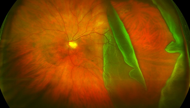
The specialist photography of the back of the eye consists of an adapted microscope connected to a flash camera that provides a vertical, magnified view. A classic camera provides a view of 30 and 50° of the retinal area –with a 2.5x zoom–, with the central retina, the macula and the optic disc being the main structures that can be analysed.
There is also another form of retinography called wide field, which captures more than 200°. This means that 80% of the retina in a panoramic image that, as well as being able to view the aforementioned structures, captures a complete image of the peripheral retina.
How is it performed?
Central retinography requires prior dilation of the pupil with eye drops that take effect within around 15 minutes and lead to blurred vision and dazzle for a few hours. Wide-field retinography does not require the use of mydriatic eye drops.
Both technologies provide a very fast test that is performed in the consulting room in 10 minutes in the case of the form and less than 5 in the case of the latter, as only one photograph is required that is taken at a capture speed of 0.25 seconds.
5 keys to the retinography
- It is a painless test, there is no direct contact with the eye.
- The only associated discomfort is that the pupil must be dilated beforehand (eye drops that take 15-20 minutes to take effect) in central retinographies.
- No pupil dilation is required in wide-field retinography.
- Contact lenses do not have to be removed for any time beforehand.
- Very fast test: between 5 and 10 minutes per eye, depending on the type of retinography.
In what cases is it used?
Retinography is useful in the observation and recording of all types of disease involving the posterior pole of the eye, such as AMD, diabetic retinopathy, retinal detachment, uveitis, tumours, or retinal dystrophies such as retinitis pigmentosa, among other retinal diseases. It also provides a view of damage to the layer of nerve fibres in the retina and the optic nerve head in patients with glaucoma.
Associated treatments
You may be interested in
IMO Institute of Ocular Microsurgery
Josep María Lladó, 3
08035 Barcelona
Phone: (+34) 934 000 700
E-mail: international@imo.es
See map on Google Maps
By car
GPS navigator coordinates:
41º 24’ 38” N – 02º 07’ 29” E
Exit 7 of the Ronda de Dalt (mountain side). The clinic has a car park with more than 200 parking spaces.
By bus
Autobus H2: Rotonda de Bellesguard, parada 1540
Autobus 196: Josep Maria Lladó-Bellesguard, parada 3191
Autobuses H2, 123, 196: Ronda de Dalt – Bellesguard, parada 0071
How to arrive at IMO from:
IMO Madrid
C/ Valle de Pinares Llanos, 3
28035 Madrid
Phone: (+34) 910 783 783
See map in Google Maps
Public transport
Metro Lacoma (líne 7)
Autobuses:
- Lines 49 & 64, stop “Senda del Infante”
- Line N21, stop “Metro Lacoma”
Timetables
Patient care:
Monday to Friday, 8 a.m. to 8 p.m.
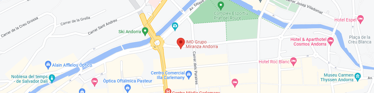
IMO Andorra
Av. de les Nacions Unides, 17
AD700 Escaldes-Engordany, Andorra
Phone: (+376) 688 55 44
See map in Google Maps
IMO Manresa
C/ Carrasco i Formiguera, 33 (Baixos)
08242 – Manresa
Tel: (+34) 938 749 160
See map in Google Maps
Public transport
FGC. Line R5 & R50 direction Manresa. Station/Stop: Baixador de Manresa
Timetables
Monday to Friday, 08:30 A.M – 13:30 PM / 15:00 PM – 20:00 PM